

Onion Cell Under Microscope 10x
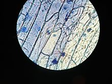
A wet mount of the onion peel nether the microscope stained with methylene bluish at 50X zoom

Fig. 1
dye, pin, onion membrane, slide, and cover slip

Fig. 2
A simple student'south microscope
Tissue from an onion is a good beginning exercise in using the microscope and viewing establish cells. The cells are easily visible nether a microscope and the training of a thin section is straight forward. An onion is made of layers, each separated by a thin skin or membrane. In this exercise, you volition make a wet mount on a microscope slide and look at the cells of the onion membrane magnified by the high power, compound microscope.
Method [edit | edit source]
- First add together a few drops of h2o or solution on the microscope slide to avert dryness and wilting
- Take a small slice of onion and using tweezers, pare off the membrane from the underside (the rough side).
- Place the membrane flat on the surface of the slide.
- Add a drop of Iodine solution to the onion pare
- Using a pivot, lower the thin glass comprehend slip or cover glass onto the slide. Brand sure there are no air bubbles (Fig. 1).
- Brand certain the everyman power objective lens (the shortest lens if there are several present) is in line with the optical tube, and the microscope light is turned on. Then place the prepared slide onto the stage of the microscope.
- Looking from the side (NOT through the eyepiece), lower the tube using the coarse focus knob until the end of the objective lens is simply above the cover drinking glass. Exercise this carefully and so every bit not to crack the cover drinking glass (and possibly damage the objective lens).
- Now look through the eyepiece and turn ONLY the smaller, fine focusing knob to move the optical tube upwards until an image comes into focus. The cells should await something similar cadger pare.
- Swap the objective lens for a higher powered ane so that you tin see the cells at greater magnification. You lot should be able to make out a nucleus in each cell.
- Be very conscientious; these dyes can stain your peel and clothes. Could be dangerous if it is on y'all.
Proper use of the microscope - intended to prevent damage to the objective lenses - requires that the following techniques be followed:
- Never use the fibroid focus knob while looking through the eyepiece. The point of focus will exist very nigh the encompass glass. Looking from the side, lower the optical tube until the objective lens is as shut as you can go information technology to the comprehend drinking glass without really touching it. Starting with the low power objective lens is the fastest way to attain proper focus.
- Initially, slowly focus back (turn the fine focus knob to enhance the optical tube) while looking through the eyepiece. In one case the specimen comes into focus, you can make fine adjustments up or downwards with the fine focus knob without fright of damaging the slide or the microscope.
- If the specimen does not focus, raise the tube a little with the fibroid focus knob and try to focus again with the fine focus knob. Once the object is in focus, switching objective lenses should be possible without any further coarse adjustments.
Return to laboratory exercise for Chapter 2

Onion Cell Under Microscope 10x,
Source: https://en.wikibooks.org/wiki/School_Science/How_to_prepare_an_onion_cell_slide
Posted by: mossstrater.blogspot.com
0 Response to "Onion Cell Under Microscope 10x"
Post a Comment